Pterygoid muscles: Protruding the Jaw
The two pterygoid muscles are just half of the muscles that help you chew from inside your mouth, just behind your lower jaw.

Pterygoids (medial and lateral) are two of the four muscles of mastication, that is, the muscles that you use when chewing your food. The medial one is also called the internal pterygoid.
Because chewing isn’t simple closing and opening your mouth, these muscles are of great importance for the circular movements of the mandible when grinding food on one side or the other, along with the tongue that moves the food back to the grinding spot between your teeth, to either side you choose to chew onto.

These are actions that most of us don’t think about much. I hope that after reading this article, you pause while you eat and think to be grateful to those chewing muscles that help you not swallow things whole.
Pteryogoid muscles function
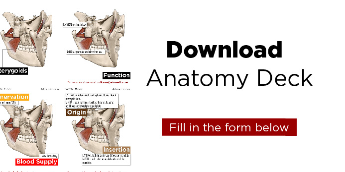
The lateral pterygoid pushes the jaw out (protrude) and also lowers it. Say, if you want to pull out your lower row of teeth and bite your upper lip, say thanks to your lateral pterygoids with it’s superior and inferior heads. Since it originates from the sphenoid bone (somewhat behind your cheeks) and inserts in the jaw (the jaw being the movable part of this arrangement and the sphenoid bone where it anchors), you can imagine how contraction of it can push the jaw out when both sides are activated. When only one side is contracted, it will move the jaw medially, side to side.
When the medial pterygoid contracts, it pushes the jaw up, closing the mouth. Because this muscle is placed diagonally, it pushes the jaw up and forward (protrusion) at the same time, as it closes the mouth. When only one side is contracted, it works with the lateral pterygoids to swing the jaw to the other side.
All these actions are really important for chewing in general, be it the obvious ones (i.e opening and closing your mouth) or the more finer ones that are less obvious (i.e. chewing on one side).
Masseter hypertrophy and pterygoids
Masseter muscle is another of the chewing muscles that work alongside pterygoids. A study found that people who are stressed and tend to clench their teeth repeatedly or grind their teeth while sleeping, over using their masseters, also have hypertrophic (bigger) pterygoids, highlighting the special connection and team work of these muscles.
And since it’s more obvious to notice a pronounced masseter (because it’s outside and makes your face look tough and square) than the pterygoids that are inside, this can prompt clinicians that notice a hypertrophic masseter to look further and extend the treatment to these.
Of the four mastication muscles, lateral pterygoids are the only ones that help opening the mouth.
Masseter muscle facts
Nerve and blood supply
Lateral and medial pterygoids muscles are innervated by branches of the mandibular division of the trigeminal nerve (VIII).
The blood supply comes from the pterygoid branches of the maxillary artery.
Muscle Attachments
Lateral
The superior head originates in the infratemporal crest of the greater wing of the sphenoid bone. The inferior head originates in the lateral surface of the lateral pterygoid plate. Both heads come together and insert into the pterygoid fovea on the neck of the mandible.
Medial
The superficial head comes from the pyramidal process of the palatine bone and the maxillary tuberosity. The deep head arises from the pyramidal process of the sphenoid bone and the medial surface of the lateral pterygoid plate. They come together at their insertion point in the triangular impression of the medial surface of the mandible.
References
Standring, Susan. Gray’s Anatomy: The Anatomical Basis of Clinical Practice. , 2016. Print.

Leave a Reply