Nasalis Muscle: flaring your nostrils

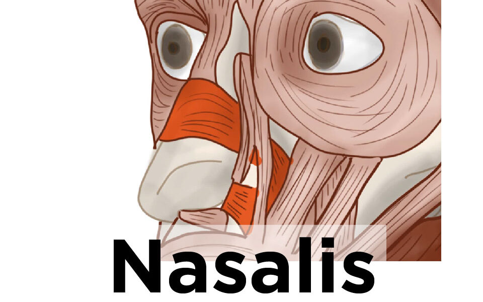
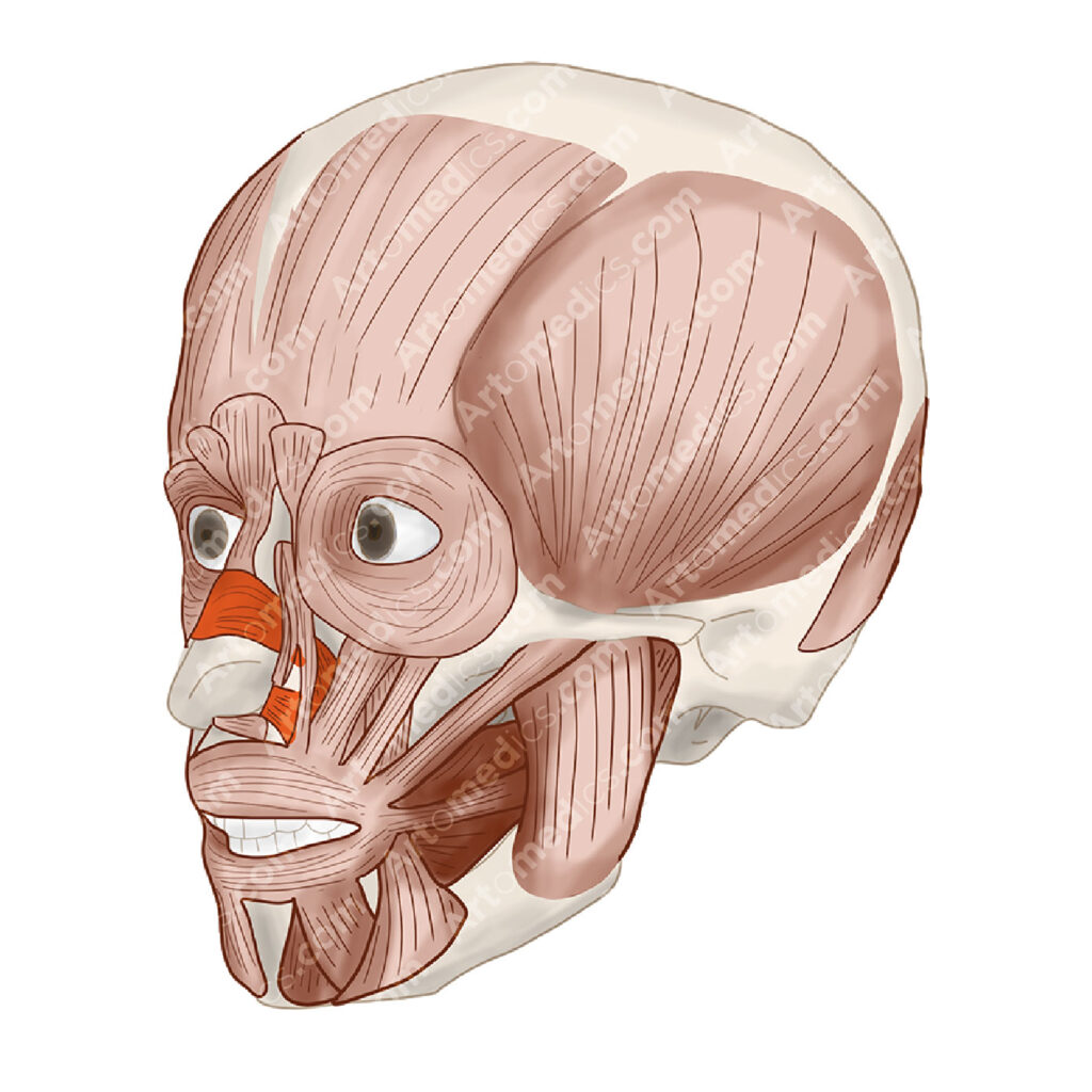
What muscle flares the nostrils? It’s the Nasalis muscle. It’s kind of sphincter with two parts, that allows you to move your nose for different facial expressions like wrinkling your nose.

Nasalis is a facial expression muscle, we use it all the time to convey certain emotions such as anger, confusion and disgust. As with all muscles of facial expression, nasalis is innervated by the facial nerve (VII), originating from the 2nd pharyngeal arch when we’re in development in the womb. This muscle is considered a sort of sphincter (like orbicularis oris and oculi) that when compressed it moves the nasal cartilages.
It is said that some people can effectively constrict it enough so that water doesn’t get into their nostrils when swimming and while underwater. I know I can’t do, but it would explain an evolutionary benefit of having our noses with muscle and enabled to move or constrict.
It’s also an important muscle to repair when approaching cleft palate surgeries, since dissecting and repairing it creates a better end-result.

What does the Nasalis Muscle do?
The nasalis muscle is generally considered in two parts: alar part (wings of the nose) and the transverse part.
Naturally, they both work in conjunction to move your nose to the sides, wrinkle it and flare up the wings. The alar part has more influence on the skin of the wings (alar means wing), lowering it to the sides and also opening the nostrils.
The transverse part wrinkles the skin on the top of your nose.
It’s a paired muscle and it moves your nose.
Nasalis Muscle Basics
Innervation
Buccal nerve. Branch of the facial nerve
Blood Supply
- Branches of the maxillary artery
- Infraorbital artery
- Branches of the Facial Artery
- Superior labial artery
- Septal artery
- Lateral nasal artery
Muscle Attachments
Muscle origin
Alar portion: it’s the part around the nostrils. It comes from the frontal process of the maxilla, just above the lateral incisor.
Transversal Portion: This part covers most of the nose. It arises from the maxilla bone, above and laterally to the incisive fossa. The fibers cross the midline of the nose, forming an aponeurosis where the bridge of the nose lies.
Muscle Insertion
Alar portion: Skin of the nose’s wing.
Transversal Portion: It’s aponeurosis in the middle of the nose blends with the other side of the muscle.
Reference
Standring, Susan. Gray’s Anatomy: The Anatomical Basis of Clinical Practice. , 2016. Print.

Leave a Reply